California

With California, Optos has incorporated new hardware and software technology enabling practitioners  to see more, discover more and effectively treat more ocular pathology thus promoting patient health.  We are committed to further strengthening our clinical evidence while demonstrating the importance of imaging the entire retina.

We have created three versions of California to be customizable for use in multiple eyecare settings.

California includes a new UWF optomap imaging modality; Indocyanine Green angiography (icg) while retaining:

  • Composite color
  • Red-free
  • Autofluorescence (af)
  • Fluorescein angiography (fa)

Images are now presented in ProView which displays optomap in a consistent geometry that accurately represents anatomical features in the retina. Further, ProView enables automatic image registration for disease tracking over time, and inter-modality image comparison.

New proprietary optical hardware optimizes and maintains resolution of the optomapimages throughout the scan of the retina resulting in more clarity in the far periphery.

Image overlay enables comparison between composite color images and red-free, af, fa, or icg images.  Additionally, comparisons can be made between different images or different dates by scrolling through all stored images.

Category:

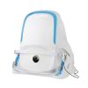
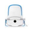
Benefits of California

In addition to the benefits found with all of the UWF devices from Optos such as 200 degrees or up to 82% of the retina captured in a single image, in multiple modalities, as well as eyecare professionals being able to see 50% more of the retina when compared to other conventional imaging devices, California offers the following benefits:

  • Compact to reduce space requirements.
  • New design leads to ease of use and faster image capture.
  • Non-mydriatic high-resolution imaging through many cataracts and/or 2mm pupils saves time in busy practices.
  • Browser-based image review enables simple integration and easy access to your data from any connected pc or tablet in a HIPAA compliant environment.
  • Interweaved angiography enables parallel capture of fa and icg images without manually switching between imaging modalities.

Technical Specifications

Image Modalities

optomap color rg and optomap plus (red and green laser):
   Color composite
   Sensory Red-free
   Choroidal
optomap color rgb (red, green, blue laser)
optomap af (green laser): autofluorescence
optomap af (blue laser): autofluorescence
optomap fa (blue laser): fluorescein angiography
optomap icg (infra-red): indocyanine green angiography 

Resolution

optomap plus: 14 μm
optomap: 20 μm

Wavelengths

Red laser: 635 nm
Green laser: 532 nm
Blue laser: 488 nm
Infrared laser: 802 nm

Exposure Time

Less than 0.4 seconds

Footprint

Width: 550 mm / 22 inches
Depth: 550 mm / 22 inches including chinrest
Height: 608-632 mm / 24-25 inches

Weight

34 kg/ 75 lbs

Laser Class

Laser safety Class-1 following IEC/EN60825-1:2007
and 21 CFR1040.10 and 1040.11

System Voltage

US: 100-120V at 50/60Hz
EU/AU: 200-240V at 50-60Hz

Power Consumption

Max. 300 VA

Communication Protocol

DICOM Compatible

Document

California – Product Brochure. Download here

Scroll to Top
×