Silverstone

Silverstone, the most powerful tool yet for examining the retina, is the only ultra-widefield imaging device with integrated swept source OCT.  Silverstone produces a 200° single-capture retinal image of unrivaled clarity in less than ½ second and enables optomap guided OCT scanning across the retina and into the far periphery.


Category:

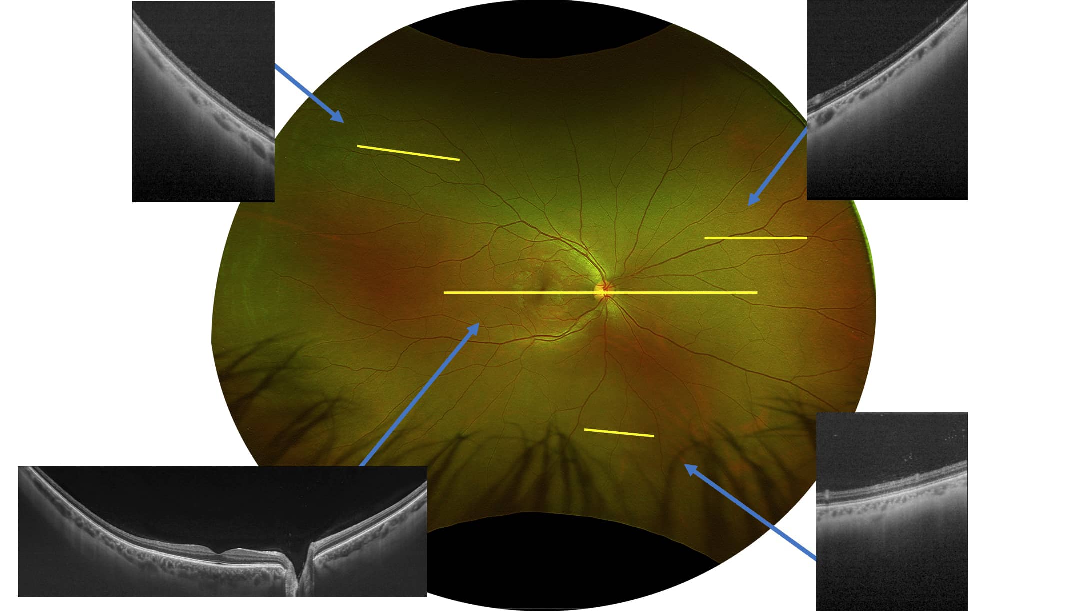
Benefits of Silverstone

  • UWF with integrated swept-source OCT, facilitates detailed examination of the retina-vitreous to sclera
  • UWF guided, swept-source OCT, images pathology anywhere on the optomap
  • 1050 nm OCT light source, provides deeper tissue penetration for clear, detailed choroidal imaging
  • 3-in-1 Color Depth ImagingTM provides important clinical data from the retinal surface through the choroid

Specification

Image Modalities optomap color and optomap plus (red and green laser):
Color composite view
Green laser view
Red laser view
optomap af (green laser): autofluorescence
optomap fa (blue laser): fluorescein angiography
optomap icg (infra-red): indocyanine green angiography
Swept Source Optical Coherence Tomography (OCT)
Resolution optomap color: 20 μm
optomap plusaf : 14 μm
Wavelengths Red laser: 635 nm
Green laser: 532 nm (for af )
Blue laser: 488 nm (for fa)
Infra-red: 802 nm (for icg)
Exposure Time Less than 0.4 seconds
Tomographic Imaging Signal Type: Optical scattering from tissue
Signal Source: Swept source OCT, Wavelength 1050 nm
Optical Power: Laser safety Class-1 following IEC/en60825-1:2014(2007)Axial Resolution: <7 micron
Transverse Resolution: <20 micron (in tissue)
Scanners: Galavanometric with x, y pair
Scan Depth: Up to 2.5mm
OCT Scan Characteristics Swept Source OCT
A-Scan rate up to 100k cycles/s
OCT Scan Types Line Scan
Volume Scan
High Density Scan
Footprint Width: 540 mm/22 inches
Depth: 570 mm/23 inches (including chin rest)
Height: 683-707 mm/27-28 inches

Image

Document

Silverstone brochure  Download

Scroll to Top
×